Elk jaar houdt The Wellcome Trust, een Engels goed doel dat biomedisch onderzoek steunt, een fotowedstrijd. Het is alleen niet zomaar een fotowedstrijd, want alle foto’s zijn gemaakt tijdens wetenschappelijke onderzoeken, of bijvoorbeeld door dokters die patienten ontvingen. De resultaten zijn vaak verbluffend, van een vrouw met een bochel, tot foto’s genomen met een elektronen-microscoop.
“De prachtige foto’s helpen ons om complexe en abstracte concepten te begrijpen,” zegt Adam Rutherfrod, één van de wetenschappers en juryleden die de fotos beoordelen.”Het gaat niet om het fotograferen van zo klein mogelijke voorwerpen, maar om het begrijpen van leven, de dood, seks en ziekte: de hoekstenen van drama en kunst.”
Twintig maart worden de winnende foto’s aangekondigd, en daarna zijn alle kandidaten te bewonderen in verschillende instituten en musea. Hieronder kan je alvast alle finalisten bewonderen, samen met de korte officiële beschrijving van wat je ziet, want dat maakt de foto’s vaak nog fascinerender. Kijk en geniet! (Tip: klik op de foto’s om ze te vergroten en in hun volledige resolutie te bewonderen)
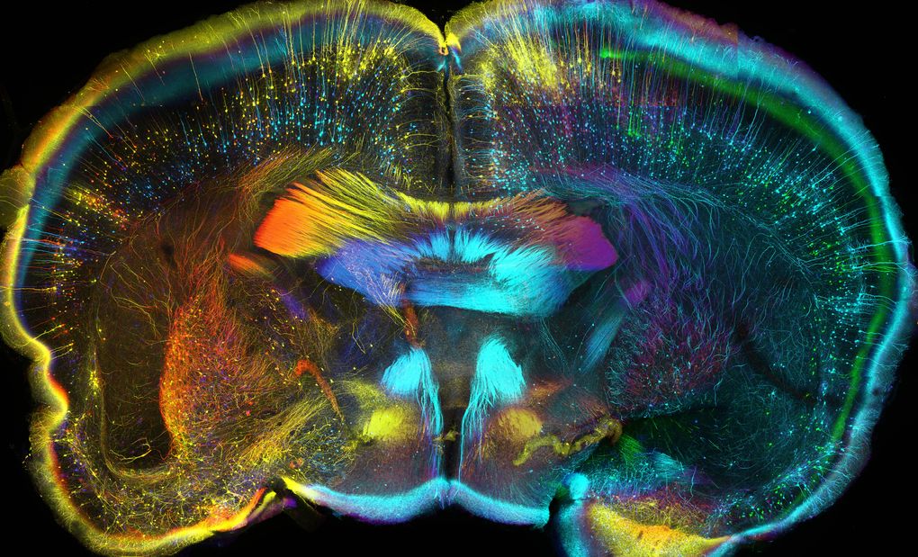
This confocal micrograph shows the nerves in a cross-section of an adult mouse’s brain. (Image credit: Luis de la Torre-Ubieta)
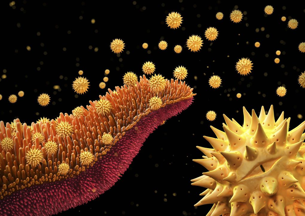
An illustration of grains of pollen being released from a a flower in the Asteraceae family. (Image credit: Maurizio De Angelis)
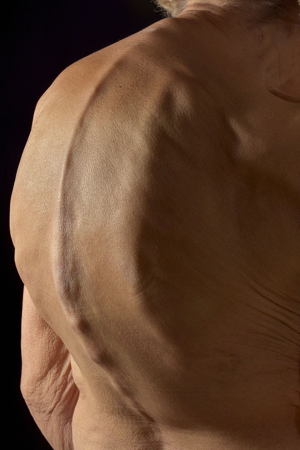
This photograph shows the spine of a 79-year-old woman with kyphosis, or “dowager’s hump.” (Image credit: Mark Bartley)
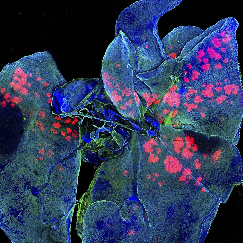
This image shows the lungs of a mouse, with the pink spots used to indicate microparticles being delivered to the lungs to research cancer medicine. (Image credit: Gregory Szeto, Adelaide Tovar, and Jeffrey Wyckoff)
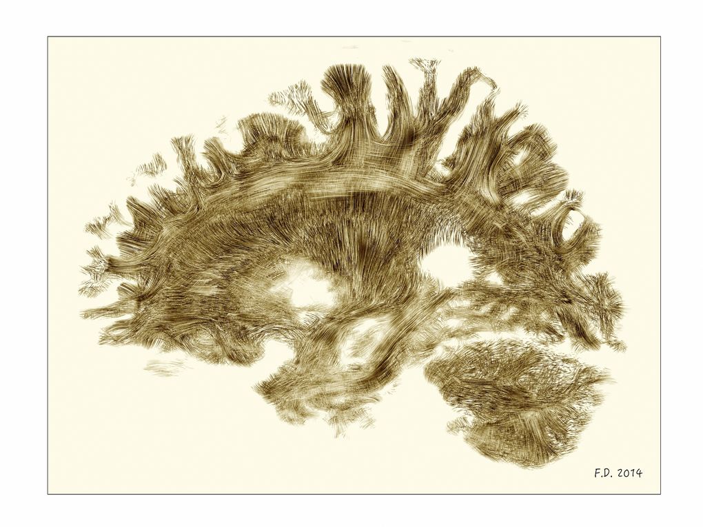
An image in the style of the 19th Century drawings of the French neurologist Joseph Jules Dejerine. It shows an MRI of a healthy, adult human brain. Image credit: Dr Flavio Dell’Acqua.
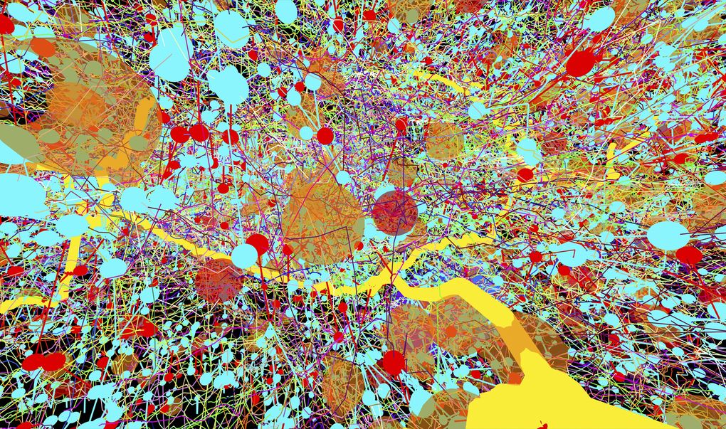
Transmission electron micrographs were used to create this color-coded representation of a fruit-fly’s nervous system. Image credit: Albert Cardona.
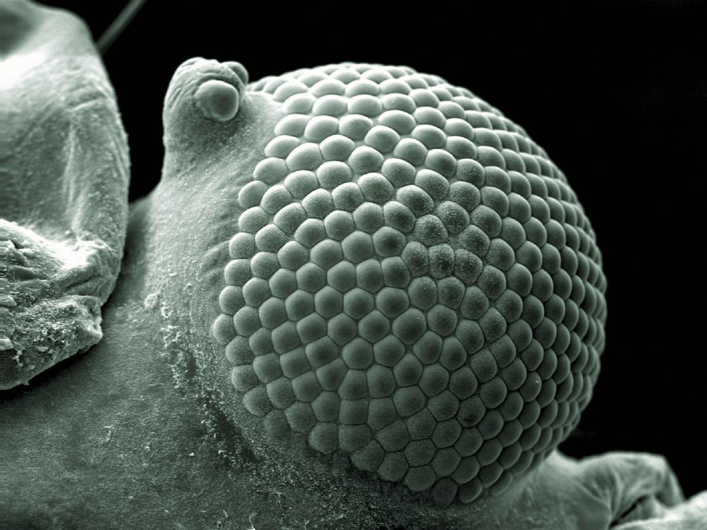
Each of these bumps contain lenses, which work together to form a greenfly’s eye. It can spot quick movements, but can’t see far. Image credit: Kevin Mackenzie.
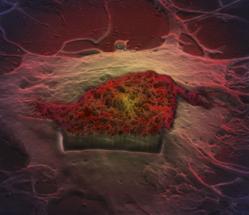
A scanning electron micrograph of a brain cell with a rectangular hole cut in it. Image credit: Khuloud T Al-Jamal, Serene Tay, and Michael Cicirko.
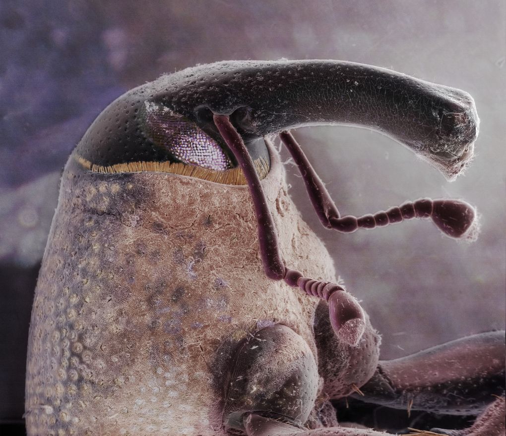
This scanning electron image shows the head of a boll weevil. (Image credit: Daniel Kariko)
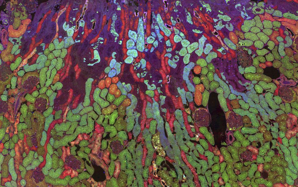
The colors in this image show chemical reactions in part of a mouse kidney. (Image credit: Jefferson R Brown, Robert E Marc, Bryan W Jones, Glen Prusky and Nazia Alam)
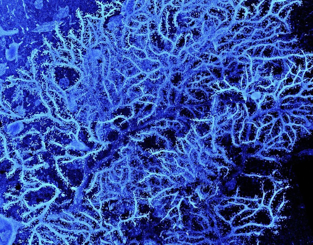
This ion beam scanning electron micrograph shows the coral-like branches of a Purkinje cell, or neuron. Found in the brain of a rat. (Image credit: Prof M Hausser, Sarah Rieubland, and Arnd Roth)
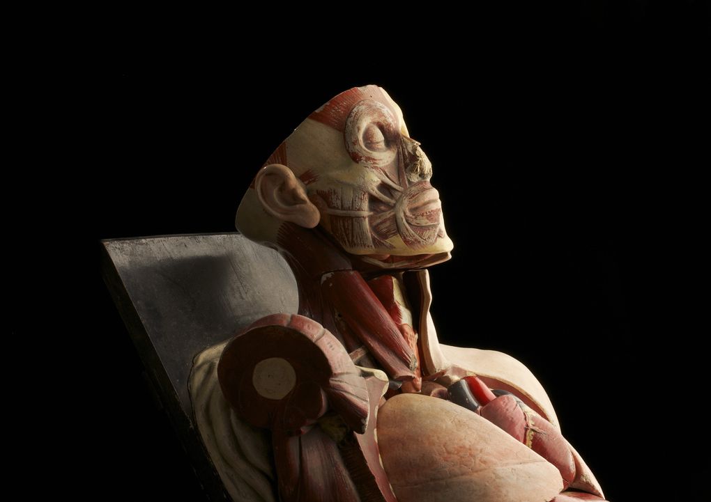
An old anatomy model. It was about to be thrown out from Trinity College in Dublin before it was rescued by the photographer. (Image credit: Anthony Edwards)
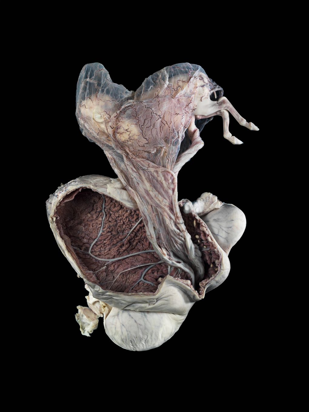
The uterus of a pony with a five month old fetus. The pony was killed in England as part of a regular cull of ponies in the New Forest. (Image credit: Michael Frank)
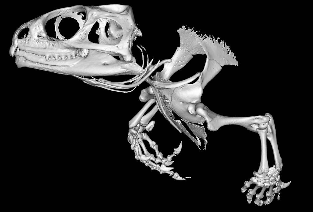
This is a micro-computed tomography (micro-CT) scan of the skull and front legs of a Tuatara — a reptile native to New Zealand with the characteristics of both lizards and amphibians. (Image credit: Sophie Regnault)
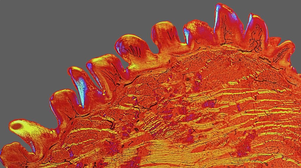
The surface of a cat’s tongue from an old Victorian slide. It shows the rough texture of the tongue. (Image credit: David Linstead)
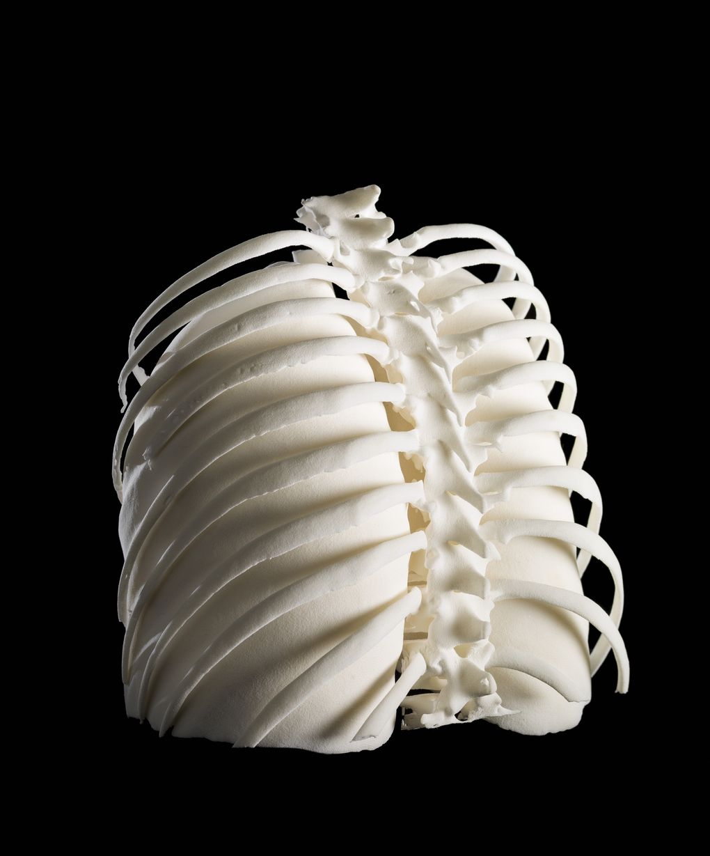
A 3D-printed model of the back of the ribcage and lungs of a woman diagnosed with cancer. (Image credit: Dave Farnham)
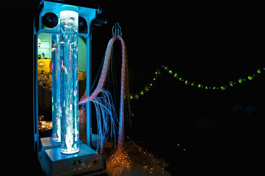
An interactive multi-sensory unit used to distract nervous or anxious children undergoing treatment in hospitals. (Image credit: Geraldine Thompson)
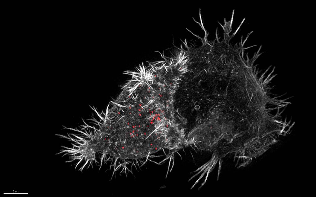
A super-resolution micrograph shows an NK cell (the pointed mass on the left) wrapping around a second cell (the slightly less pointy mass on the right). (Image credit: N Dieckmann and N Lawrence)
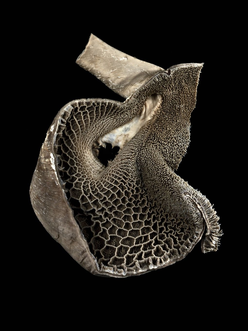
This photograph shows the interior of a goat’s reticulum — the second of its four stomachs. (Image credit: Michael Frank)
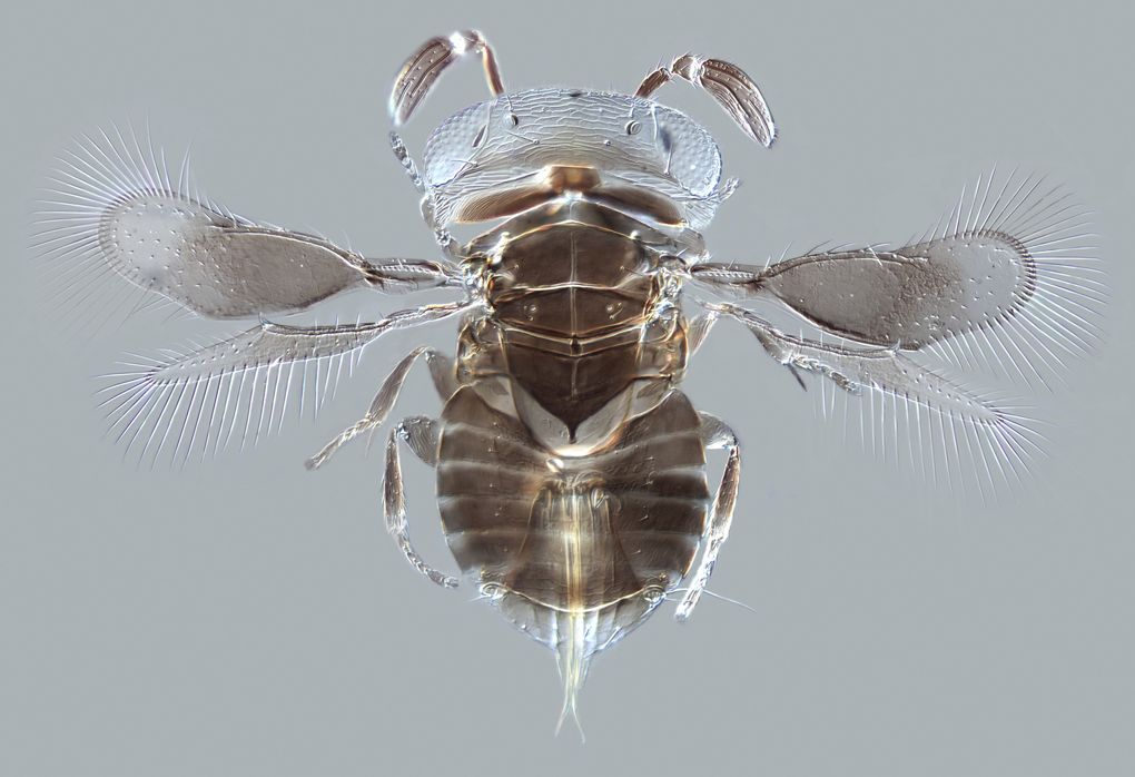
A tiny parasite wasp that lays its eggs in other animals. It’s just .75 millimeters long. (Image credit: Andrew Polaszek)